|
|
|
WorkshopsFIMH 2023 will feature 4 satellite events, detailed below. We remind you that it is necessary to register to be able to attend these different workshops. Information on the registration fees is detailed on the Registration page. ______________________________________________________________________________________________ MYOSAIQ: challenge on MYOcardial Segmentation with Automated Infarct Quantification
Date: Monday, June 19th (9am to 12am) Organizing Committee: Olivier Bernard, Magalie Viallon, Pierre Croisille, Nicolas Duchateau, Patrick Clarysse, Lucas Braz, William Romero, Frederic Cervenansky Program: The clinical use of late gadolinium enhanced magnetic resonance images (LGE) involves quantifying the areas of myocardial muscle affected by a myocardial infarction. To this end, this challenge aims to accomplish two objectives: (i) compare the performance of automatic methods for segmenting the left ventricular endocardium and epicardium on LGE images; (ii) compare the performance of automatic methods for quantifying the size of myocardial infarction lesions at different phases of the disease's longitudinal evolution, using images acquired at the acute phase (4-8 days post-MI and reperfusion therapy) and delayed phase (1 month / 12 months post-MI and reperfusion). The originality of this challenge lies in the analysis of a large database composed of 467 MR-LGE exams and integrating the main various issues encountered in daily clinical cardiac imaging, namely: the longitudinal evolution of the patho-physiology of the heart, the presence of image artifacts, the use of different MR acquisition techniques (3D versus 2D PSIR), the use of different MR acquisition settings from different centers. More information can be found on the challenge website.
______________________________________________________________________________________________ Cardiac Kinematics Benchmark
Date: Monday, June 19th (1.30pm to 5pm) Organizing Committee:
Background, motivation, and scope: Cardiac strains are a promising biomarker of cardiac health and have the potential to guide diagnosis, prognosis, and therapy planning. However, to date, cardiac strains are not routinely used in the clinical setting. One of the main obstacles to incorporating cardiac strains in the clinical workflow is the lack of accepted quantification standards and validation criteria. The goal of this workshop is to continue the community-wide discussion initiated at FIMH 2021 on establishing standards, requirements, and validation criteria that are necessary to promote the future routine use of cardiac strains in clinical practice. We have invited four experts in the field to discuss the following
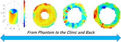
In addition, we will present an update on the findings of the Cardiac Kinematics Benchmark held at FIMH 2021 to drive discussions around cardiac strains variability, reproducibility, and uncertainty in a practical test case. The long-term goal of this workshop is to form an international working group to advance the field of cardiac kinematics, and we hope this year's discussion will contribute to improving existing validation and analysis approaches. Agenda:
______________________________________________________________________________________________ Simulation and Imaging for Mitral RegurgitationDate: Thursday, June 22th (1.30pm to 5pm) Organising Committee: Miguel Fernandez Background, motivation, and scope: Mitral Valve regurgitation, also known as mitral insufficiency, is one of the most important cardiac valve disease. Primary mitral valve regurgitation often manifests with elongation or rupture of chordae leading to prolapse. The repair of the vale via “respect” approaches which preserve the leaflet tissue, i.e, implantation of artificial chordae (neochordae) or clips (MitraClip), has shown very good results. Nevertheless, one of the main issues in these techniques is the lack of bio-physical insight on the repair (which neochordae length?, which chordae tension?, which coaptation forces?, etc.). Echocardiography is the most common tool to study the mitral valve regurgitation, but this imaging modality is sometimes insufficient to determine the optimal timing for surgery, identify the best surgical strategy and predict early failures after surgical repairs. New quantitative measurement tools are hence needed to understand the bio-physical consequences of mitral valve repair. The aim of this workshop is to provide a forum for discussions and exchanges on the clinical, imaging, experimental and simulation aspects of this problem. This workshop will be held in the framework of the ANR SIMR project (2020-2024) and hosted by the FIMH conference in Lyon. Program:
______________________________________________________________________________________________ Simulation of echocardiographic sequences (hands-on)
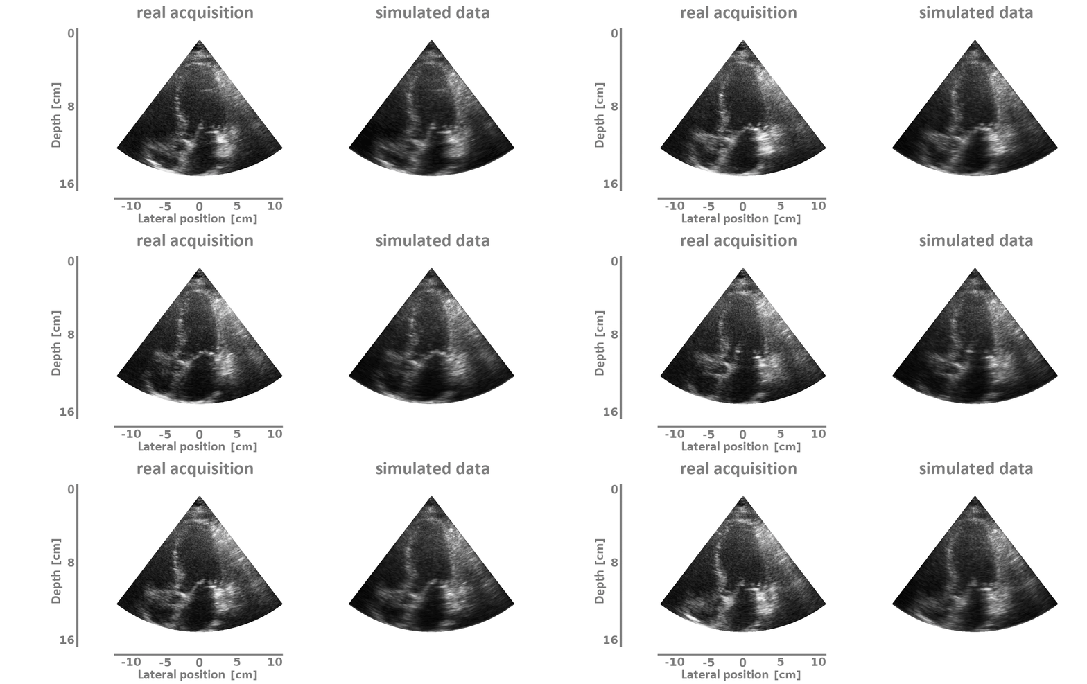
Date: Thursday, June 22th (1.30pm to 5pm) Organizing Committee: Damien Garcia, Olivier Bernard Localization: Gustave Ferrié building - 1st floor - 6 Rue de la Physique, Campus de la Doua - 10 minutes walk - google map Program: Echocardiography is a valuable diagnostic tool for assessing cardiac function and diagnosing heart disease. However, acquiring expertise in this imaging technique requires extensive training and experience. In recent years, the use of artificial intelligence, specifically deep learning neural networks, has shown promise in improving the accuracy and efficiency of echocardiographic image interpretation. Realistic simulations of echocardiographic images can play a crucial role in training and testing these neural networks. This workshop will introduce SIMUS, an ultrasound image simulator developed in Matlab that can generate realistic echocardiographic images for training deep learning neural networks. The goal of this workshop is to provide participants with hands-on experience in using SIMUS to generate realistic echocardiographic images as given below.
|


